This page is lifted from Sir V’s handout on Perioperative Nursing. He has a lot more to say than what is on the presentation, so don’t expect this to be completely comprehensive in terms of content. This can be used as a content outline for further review. However, I did attempt to supplement information when possible to provide more context.
When I have time, full lectures on perioperative nursing will be linked here. The sources I have for perioperative nursing are all pretty long, so they will have to wait.
Perioperative Nursing
Perioperative nursing is the identification of the physiological, psychological, social and spiritual needs of the client and the formulation of an individualized plan of care before, during, and after surgery. Surgeries are done to cure, to relieve pain, to prolong life, to maintain dynamic body equilibrium, to treat and prevent infection, or to correct deformities or defects. For this vast array of functions, surgeries take on various forms:
| Classification | Types |
|---|---|
| According to Purpose | - Curative: to cure - Diagnostic: to diagnose - Exploratory: to explore |
| According to Location | - External - Internal |
| According to Mode | - Constructive: to create structures - Reconstructive: to restore structures |
| According to Degree of Risk | - Minor - Major |
| According to Urgency | - Emergency - Urgent - Elective: may still be required, but can be scheduled/voluntary. |
The Surgical Team
The medical team handling an patient in the operating room is composed to a multidisciplinary set of members that all play their respective roles for providing holistic care. They are generally divided between sterile and unsterile members of the team:
| Team | Members |
|---|---|
| Sterile | 1. Operating Surgeon 2. Assisting Surgeons 3. Scrub Nurse |
| Unsterile | 4. Anesthesiologist 5. Circulating Nurse |
Scrub Nurse
- Receives patient from the surgical ward nurse.
- Prepares and organizes the OR unit based on the case.
- Opens sterile packs and adds sterile supplies and instruments.
- Performs surgical scrubbing, gowning, and gloving.
- Organizes sterile fields.
- Serves gowns and gloves to surgeons.
- Counting of instruments.
- Draping.
- Announcing cutting time.
Circulating Nurse
- Receives patient from the surgical ward nurse. Endorsement during transfer includes confirmation of patient identity, schedule, and securing informed consent. Discussed later: Preoperative Preparation
- Establishes rapport with the client.
- Places the patient on the OR table and remains with them.
- Position the patient for anesthesia. This may be supine or quasi-fetal position depending on the anesthesia being used.
- Perform lumbar preparation for spinal or epidural anesthesia.
- Catheterize the patient if necessary.
- Skin preparation.
- Position the patient for surgery.
- Help drape the patient.
- Record cutting time.
Surgical Landmarks and Positioning
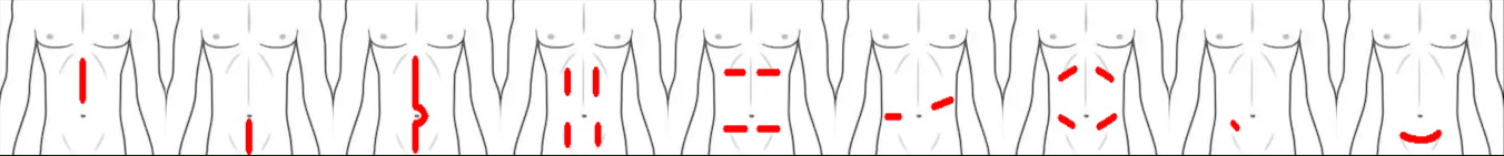
Many landmarks and subdivisions are used in surgery, used appropriately depending on the purpose of the surgery. Surgical incisions include:
- Upper Midline Incision: midline incision above the umbilicus
- Lower Midline Incision: midline incision below the umbilicus
- Longitudinal Midline Incision: midline incision above, around, and below the umbilicus. Not through the umbilicus directly.
- Paramedian Incision: meaning “adjacent to the midline”. It is a longitudinal incision performed in one or more of the four quadrants of the abdomen.
- Mid-abdominal Transverse Incisions: same as paramedian incisions, but perpendicular to the long axis of the body.
- Thoracolumbar Incisions (no clue…)
- Oblique Incisions: incisions similar to paramedian incisions, but at oblique angles.
- McBurney’s Incisions: an incision at McBurney’s point, the midpoint between the umbilicus and the right anterosuperior iliac spine. This is used to locate the position of the appendix, and is the most common site of maximum tenderness in acute appendicitis.
- Pfannenstiel Incisions: the most common method for performing caesarian sections in present day. It is a slightly curved transverse incision between the symphysis pubis and umbilicus.
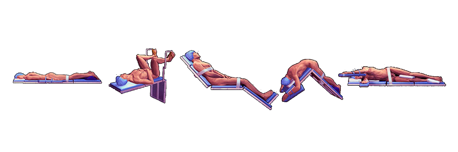
The patient is positioned in whichever position is most ideal for the area being operated on. This includes:
- Prone Position: the patient lies on their stomach with their head turned to one side. This is used for spinal, rectal, or perianal procedures.
- Lithotomy Position: the patient is supine with their hips and knees flexed, with the legs elevated and supported by stirrups. This is used for gynecological, urological, and colorectal procedures.
- Semi-Fowler’s Position: supine, with the head of the bed elevated at 30° to 45°. This is used for head, neck, or chest procedures.
- Kraske/Jackknife Position: the patient is positioned prone, with the operating table set to flex at the hip. This elevates the pelvis above the rest of the body. This is used for rectal and some spinal procedures.
- Lateral Position: the patient is positioned on their side (side-lying). This is used for thoracic, renal, and hip procedures. Proper padding and positioning aids is required for proper alignment and stability.
- Trendelenburg Position: a normal supine position, with the head of the bed lowered and foot of the bed raised. The body maintains a neutral and straight posture.
- Reverse Trendelenburg Position: the Trendelenburg position with the head elevated.
- Kidney Position: an amalgamation of the jackknife position and the lateral position. The patient is side-lying, bent at the level of the kidney (around the costovertebral angle) to make it protrude above the rest of the body.
The abdominal cavity is occupied by many organs of the gastrointestinal system, renal system, reproductive systems (females), etc. There are two general subdivisions of the abdomen:
- Nine-Region System: this system divides the abdomen into nine areas using two horizontal and two vertical lines (midclavicular lines). The lowest region ends at the level of the highest part of the pelvic bone (transtubercular line), and the highest region ends at the end of the ribcage (subcostal line).
| Left | Middle | Right |
|---|---|---|
| Left Hypochondriac | Epigastric | Right Hypochondriac |
| Left Lumbar | Umbilical | Right Lumbar |
| Left Iliac (Inguinal) | Hypogastric (Suprapubic) | Right Iliac (Inguinal) |
- Four-Quadrant System: this system divides the abdomen into four with the midline from the xiphoid to the symphysis pubis, and a transverse cut across the umbilicus.
| Left | Right |
|---|---|
| Left Upper Quadrant | Right Upper Quadrant |
| Left Lower Quadrant | Right Lower Quadrant |
Preoperative Phase
The patient has to undergo preparations prior to surgery, with some requiring preparation days or weeks prior to surgery. Generally, these are the preoperative preparations to perform the day and night before the surgery:
- Pre-operative visit the day before the surgery.
- Client education on post-operative activities, such as breathing exercises to avoid pulmonary atelectasis, splinting coughs, etc.
- Complete laboratory and diagnostic examination coordinated with the MD.
- Cardiopulmonary (CP) Clearance: an initial evaluation to determine if the patient will be able to undergo surgical intervention on general anesthesia. Not being cleared indicates an elevated risk for cardiac events during surgery.
- Checking of blood products when indicated.
- Monitoring VS and I&O.
- Secure consent.
- Bathing as necessary.
- Light evening meal and NPO from midnight to the time of the surgery. No oral intake should be had to prevent emesis and pulmonary aspiration of gastric contents when placed under general anesthesia. Failure to maintain NPO status can be grounds for delaying surgery.
- Psychological and spiritual support.
- Administration of laxatives if indicated.
- Removal of nail polish (for accurate pulse oximetry).
On the morning of the surgery itself, the most important nursing actions include:
- Ensuring NPO for reasons previously mentioned.
- Oral care.
- Enema if ordered.
- Shaving areas to be operated on.
- Reviewing post-op exercises like those previously mentioned.
- Preoperative medications are also given at this time. They are discussed later in this section.
- Monitoring of patient status, including their psychological state. Provide support as necessary.
- Removal of dentures as aspiration precautions.
- Endorsement to the OR from the surgical ward.
Pre-operative medications are given to prepare the client for anesthesia and potentiate its effects, and to sedate the client to reduce anxiety levels. These include:
- Narcotic Analgesia
- Sedatives
- Anticholinergics
Consent
Informed Consent is a condition of understanding and willingness to undergo a procedure. The distinction of “Informed” specifies that the patient understands all pertinent information related to the procedure, such as purpose, risks, benefits, alternatives, side effects, cost, among others. For this, the patient is required to be of sound mind and of age.
Also read: Consent in Health Care
Intraoperative Phase
The intraoperative phase includes the goals of care of asepsis, infection control, homeostasis, safe administration of anesthesia, and hemostasis.
Asepsis
Asepsis, disinfection, and sterilization are all varying ways to reduce or completely eliminate the presence of microorganisms on a field or body to produce an environment viable for surgical procedure.
- Asepsis: the elimination of microorganisms from living tissue.
- Disinfection: the elimination of microorganisms from inanimate objects, excluding spores.
- This may be done through physical or chemical methods. Physical methods include boiling, steaming, and sunlight. Chemical methods include alcohol, chlorine, iodine, and phenol.
- Sterilization: the elimination of microorganisms from inanimate objects, including spores.
- This may be done through physical or chemical methods. Physical methods include autoclaving, the main method of sterilization, radiation, and gas. Chemical methods mainly involve soaking/immersion.
// skipped “Sample Sterilization and Level of Disinfection for Glutaradehyde”
Earle Spaulding created the Spaulding Classification to determine the level of disinfection required for items being used for
- Critical: used for items that cut into intact skin and mucous membranes or items entering vascular areas of the body. This requires sterilization.
- Semi-Critical: used for items coming into contact with non-intact skin and mucous membranes. This requires high-level disinfection.
- Non-Critical: used for items coming into contact with intact skin and mucous membranes. This requires intermediate-level to low-level disinfection.
Principles of Sterile Technique
- Only sterile items are used within the sterile field.
- Sterile personnel are gowned and gloved.
- Tables are sterile only at table level.
- Sterile personnel touch only sterile items or areas; unsterile personnel touch only unsterile items or areas.
- Unsterile personnel avoid reaching over the sterile field and sterile personnel avoid leaning over an unsterile area.
- The edges of anything that encloses sterile contents are considered unsterile.
- The sterile field is created as close as possible to the time of use.
- Sterile areas are continuously kept in view.
- Sterile personnel keep well within the sterile area.
- Sterile personnel keep contact with sterile areas to a minimum.
- Destruction of the integrity of microbial barriers results in contamination.
Surgical Scrubbing
| Time Method | 1st (5 min.) | 2nd (4 min.) | 3rd (1 min.) |
|---|---|---|---|
| Hand | 1 minute | 1 minute | 30 seconds |
| Arm | 1 minute | 1 minute | None |
| Elbow | 30 seconds | None | None |
| Brush-Stroke Method | 1st (5 min.) | 2nd (4 min.) | 3rd (1 min.) |
|---|---|---|---|
| Fingertips | 10/3 | 5/3 | 3 |
| Hand | 10 | 5 | 3 |
| Arm | 6 | 3 | 0 |
| Elbow | 6 | 0 | 0 |
Surgical Instrumentation
Instruments used during surgeries are generally divided between sharps to cut, graspers to hold, clamps to occlude, and retractors to reveal.
I really can’t be bothered to add images for all of these. Use Google if you’re not familiar with them.
Sharps
Equipment used for cutting, dissecting, or piercing tissues.
- Knives: the primary cutting tool for membranes. These come in differing sizes for varying tissues. Blades from 20 to 25 are paired with handle #4, and is used to cut tough tissues. Blades from 10 to 15 are paired with handle #3, and is used to cut delicate tissues.
- Handles: the handle for knives. These are either #4 for blades 20, 21, 22, 23 and 25, or #3 for blades 10, 11, 12, 13, and 15.
- Only when a knife is placed on a handle is a scalpel formed.
- Scissors:
- Curved Mayo Scissors: used for heavy or tough tissues.
- Metzenbaum (Metz) Scissors: used for delicate tissues.
- Straight Scissors: used for cutting sutures.
- Bandage Scissors: cutting tough tissues, particularly the uterus and the umbilical cord during caesarean section.
- Steven’s Scissors: cutting off eyelashes
- Needles: straight or curved (cutting or round) needles used for puncturing the skin to create stitches. These are paired with ties for suturing.
- Sutures are surgical-use threads used for closing wounds/creating stitches. They may be absorbable (dissolves by itself within the body), or non-absorbable.
- Absorbable: non-synthetic— catgut, chromic, plain sutures or synthetic— Dexon, Vicryl, PDS
- Non-Absorbable: non-synthetic— silk, cotton, or synthetic— nylon.
- Sutures are surgical-use threads used for closing wounds/creating stitches. They may be absorbable (dissolves by itself within the body), or non-absorbable.
Graspers
“Grasps” tissues.
- Thumb Forceps: used to hold delicate tissues. The tip of these are toothless.
- Tissue Forceps: visually the same as thumb forceps, but feature a tooth at the end of the forceps.
- Babcock Forceps: forceps with a cylindrical cavity used to handle tubular, delicate tissues.
- Allis Forceps: toothed, rake-like forceps used for tougher tissues like bones, tendons, and fascia.
- Adson Forceps: toothed forceps used for tougher tissues.
- Pennington Forceps: triangular forceps used for perineal or rectal surgery.
- Ovum Forceps: used for delicate forceps. These are also used as pick-up forceps.
Clamps
Tools used for occluding vessels or to produce hemostasis. Each of these have straight and curved variants.
- Mosquito: the shortest clamp; used for minor surgery, pediatrics, and handling superficial layers.
- Crile: medium-sized; used for shallow layers.
- Kelly: the longest; used for abdominal layers or cavities.
- Ochsner: strong, fully-serrated clamp with a tooth at the end that crushes tissue to prevent bleeding. It is commonly used for hysterectomies.
- Mixter: a forcep that curves at the end, used to maneuver around structures to reach hard places.
Retractors
These are further specified as self-retaining (does not require manual application of force) or non-self-retaining (requires handling).
Self-Retaining
- Balfour Abdominal Retractor: a large self-retaining retractor primarily used in abdominal surgeries. Two lateral blades and a central blade can be adjusted to keep the abdominal wall open.
- Weitlaner Retractor: a small self-retaining retractor with two arms ending in outward-pointing, curved prongs.
- Mastoid Self-Retaining Retractor: a scissor-like tool with rake-like ends used for small operative areas.
- Gelpi: a scissor-like tool with pointed tips used for perineal surgery.
Non-Self-Retaining
- Army Navy Retractor: a double-edged handheld retractor with a straight handle and two blades of different lengths on each end. These are used for superficial layers.
- Richardson Retractor: a single or double-ended handheld retractor with a right-angled blade. It is a curved, concave blade that comes in different sizes.
- Deaver Retractor: a long, flat handheld retractor with a scoop-like blade at one end. It often holds back large organs like the liver or intestines, providing exposure to deep areas of the abdominal cavity.
- Bladder Retractor: a specialized handheld retractor that gently moves and holds the bladder or other pelvic organs away from the surgical site.
- Murphy/Rake Retractor: a rake-like retractor with varying number of teeth.
- Senn Retractor: also a rake-like retractor, used for superficial/skin retraction. It can be double-ended, or have one end that is curved and concave.
- Malleable Retractor: a softer retractor that can be shaped as necessary.
Sedation
Anesthesia is the loss of sensibility to pain. In administration, there are four stages of anesthesia:
- Induction: the patient feels drowsy or dizzy. Sensation of pain begins to dull.
- Excitement: breathing becomes irregular, and involuntary motor movements may appear.
- Surgical Anesthesia: the period appropriate for surgery; muscles are relaxed, pupils are constricted, and the pupil reflex is absent.
- Medullary Depression: oversedation; depression of the medulla, which can cause respiratory cessation and potentially death.
Depending on the scope of the anesthesia, it can be considered as general anesthesia (IV, inhalation) that produces sensory, motor, reflex, and mental blocks. For anesthesia that only affects one area of the body, they are considered as regional anesthesia (spinal, epidural, nerve blocks, infiltration, application, spray). The following are commonly used anesthetics:
- Non-Halogenated Inhalants:
- Nitrous Oxide (Blue) used for initial restlessness
- Cyclopropane (Orange) used for short procedures
- Halogenated Fluid Inhalants:
- Halothane (Red) used for hypotension
- Enflurane (Yellow) used for muscle relaxation
- Sevoflurane (Sweet-tasting) used for pediatric patients
- Intravenous Barbiturates: Thiopental
- Neuroleptic Agents: Fentanyl, which decreases motor activity
- Dissociative Agents: Ketamine, which can cause hallucinations
Local anesthesia is a class of anesthesia which function at a narrower scope than even regional anesthesia. These do not produce a change in the level of consciousness. These also take on different forms:
- Topical: applied over the surgical site, e.g., Lidocaine, Prilocaine (EMLA Cream)
- Field/Nerve Block: injected into subcutaneous or perineural spaces near or around the desired anesthesia site, blocking pain impulses from being felt.
- Spinal: anesthesia injected directly into the subarachnoid space (inside the arachnoid).
- Epidural: anesthesia injected into the epidural space (outside of the arachnoid), commonly used in obstetrics.
Hemostasis
Hemostasis is the cessation of hemorrhaging from an injured blood vessel. In a normal wound, this occurs in three stages: vasoconstriction, platelet plug formation, and coagulation. However, in a surgery, artificial methods can be used to induce hemostasis:
- Mechanical Pressure: manual, digital, dressing, clamping, gel foam, Penrose drain
- Chemical: tissue adhesion
- Thermal: cautery
Postoperative Phase
Immediate Care
This is care provided while the patient remains in the recovery room.
- Assure ABCs:
- O₂ therapy with positioning, artificial airways until the return of gag reflex, suctioning, encouraging deep breathing
- Check vital signs every 15 minutes until stable, then every 30 minutes.
- Check skin color, temperature, drainage, and dressings. These can
- Monitor Level of Consciousness: reorient the client. Sedation can last for a few hours and affect cognition.
- Discharge: once the patient is awake and responsive, with easy breathing and acceptable CP clearance, they may be discharged from the recovery room.
Continuing Care
Care for the postoperative client can continue in the ward. The primary concern is to promote optimal functioning, reducing pain, and wound care:
- Optimal Respiration: coughing, deep breathing, incision splinting, early ambulation, and turning in the bed are all potential exercises for maintaining and improving respiration by the patient.
- Optimal Circulation: early ambulation and leg exercises
- Optimal Nutrition
- Optimal Fluid and Electrolyte Balance: monitor IV, I&O, drains, dressings, and the return of peristalsis (flatus, bowel movement)
- Pain Control: analgesics and comfort measures
- Wound Care
Postoperative Discomforts
- Pain
- Urinary Retention, often from the use of an indwelling catheter.
- Hiccups can be a result of pathogenesis.