Reference
Salustiano, R. (2024). The Normal Antepartal Period. In Dr. RPS Maternal & Newborn Care: A Comprehensive Review Guide and Source Book for Teaching and Learning (2nd ed., pp. 80-116). C&E Publishing, Inc.
The antepartal period is the period of pregnancy or the period before labor, also called the prenatal or the antenatal period. The woman in this period is called the gravida. It is part of the perinatal period, extending from one year before to two years after (18-24 months) giving birth.
Duration of Pregnancy
A normal pregnancy lasts for 267 to 280 days; 9 calendar months; 10 lunar months; 40 weeks; or 3 trimesters. The best measurement for gestational age (AOG) and length of pregnancy is weeks. The expected date of confinement (EDC) is at 40 weeks AOG.
- First Trimester: the period of rapid organogenesis, where teratogens (alcohol, drugs, viruses, and radiation) cause the most damage.
- Second Trimester: the most comfortable period, with continued growth of the fetus.
- Third Trimester: rapid deposition of fats, iron (8th month), and calcium (7th month), as the period with the most rapid fetal growth.
Physiological Adaptations in Pregnancy
Reproductive System
The Uterus grows from 7.5 × 4 × 2.5 cm, pyriform/pear-shaped to 32 × 24 × 22 cm, globular/oval. Its weight increases from 60 ~ 70 grams to 1,000 grams. Despite this, no new uterine muscle is formed. The existing muscle fibers become hypertrophic, and new fibroelastic tissues form to make stronger uterine walls. Changes are progressive.
- 12 weeks: the corpus and fundus become globular; almost spherical.
- Fundal Height is at the level of the symphysis pubis until the 12th week, and is generally only palpable by the 13th week just above the symphysis pubis. It reaches the umbilicus by the 20th to 22nd week, and the xiphoid process by the early 36th week. This is also expressed in Bartholomew’s Rule.
- Increased vascularity (because of Estrogen) results in three probable signs of pregnancy:
- Hegar’s Sign: softening of the isthmus.
- Goodell’s Sign: softening of the cervix
- Chadwick’s Sign: blue/purple discoloration of the cervix and vaginal mucosa. This may also be seen as a presumptive sign of labor as observed by the pregnant woman.
- Braxton Hicks Contractions: intermittent, irregular, painless, abdominal, and false labor contractions felt by 4 months, and is more pronounced on the 8th month.
- Ballottement: rebounding of the fetal head against examining fingers by 4 to 5 months (16th to 18th week of gestation). This is felt by healthcare examiners on the third Leopold’s Maneuver (Pawlick’s Maneuver).
- Secondary Amenorrhea: often the first sign that alerts a woman to pregnancy; the lengthened lifespan of the corpus luteum (from two weeks to two months) prevents another menstrual period to initiate.
- Uterine Electrical Activity: low and uncoordinated in early gestation, progressively intensifying and synchronizing at term. This synchronization occurs twice as fast in multiparas.
The cervix becomes shorter, thicker, and more elastic. Its mucosal lining increases mucus production (from edema and thickening), which creates the mucus plug by week 7 that protects the uterus from bacterial contamination. As previously mentioned, this is also subject to Chadwick’s Sign, becoming discolored to blue/purple from increased vascularity. The vagina also becomes thicker and hypertrophic, thickening the vaginal mucosa. Leukorrhea occurs as whitish, mucoid, non-foul, non-pruritic vaginal secretions increase along with estrogen. Vaginal acidity increases, adding protection against bacterial invasion.
Ovulation and maturation cease as the ovaries’ function is overtaken by the corpus luteum in early pregnancy; it functions maximally during the first 6 to 7 months of pregnancy.
The perineum becomes hypertrophic, experiences edema, and relaxation. It also becomes a deeper color due to vascularization. The breasts increase in size and firmness. The areola and surrounding skin darkens and enlarges, along with the alveoli system, alveoli duct, and Montgomery’s glands. Superficial veins enlarge and become prominent. Breastfeeding can be done as early as 4 to 5 months (for other infants, if needed), as colostrum begins to be produced.
Endocrine System
The major endocrine organ during pregnancy is the placenta. Its chorion (15 to 20) secrete Human Chorionic Gonadotropins (hCG) which (a) maintain the corpus luteum by secreting progesterone in early pregnancy, its most important function, and (b) aids in diagnosing pregnancy, as it is detectable in serum (by 8 to 10 days after implantation) and urine (by 10 to 14 days after first missed menstruation). hCG is found to be elevated in pregnant women experiencing vomiting.
- The placenta matures by 10 to 12 weeks (3 months), increasing placental hormones estrogen, progesterone, hCG, and hPL/hCS (Human Placental Lactogen/Human Chorionic Somatomammotropin).
hPL is the major diabetogenic hormone in pregnancy, contributing to gestational DM.
The anterior pituitary gland becomes ready for breastfeeding through increase prolactin. FSH production from the APG does not result in ovulation. The posterior pituitary gland (PPG) secretes oxytocin from the hypothalamus when fetal head pressure on the cervix increases. Oxytocin acts to stimulate the uterine myometrium, causing uterine contractions and labor onset, especially with the decrease in progesterone, which inhibit contractions, in late pregnancy.
Thyroid changes result in increased basal metabolic rate (BMR), only returning to normal levels 6 weeks postpartum. This is due to elevated serum estrogen, placental effects, and renal clearance of iodide. Increased BMP is manifested by increased pulse rate and cardiac output, a slight rise in temperature, and heat intolerance. The Parathyroid glands enhance calcium and phosphorus metabolism to meet fetal needs of calcium. If calcium needs are not met, maternal cramping occur from calcium-phosphorus imbalance.
- The pancreas increase insulin production in response to increased metabolism.
Finally, the adrenal cortex increase cortisol production to promote metabolism and activate gluconeogenesis (protein to glucose) when energy is required, and aldosterone for sodium retention (and therefore water reabsorption), resulting in the cushingoid features of pregnancy
Respiratory Systems
The lungs have a slight increase in vital capacity. Oxygen consumption increases by 15% from 6 to 40 weeks of pregnancy. The fetus consumes oxygen and contributes carbon dioxide, demanding increased oxygen and increased excretion of carbon dioxide. This creates a tendency to hyperventilate, resulting in dizziness, lightheadedness, pallor, and tingling sensation on fingertips/lips.
- Resolve hyperventilation and avoid respiratory alkalosis by promoting re-breathing with a paper bag or cupped hands.
The gravid woman experiences increased vascularity (from estrogen) of the nose, contributing to common epistaxis, nasal stuffiness, hoarseness, eustachian tube blockage (potential temporary deafness). Respiratory rate does not vary largely, reaching a maximum of 24 respirations per minute while at rest (normally 20). Lung volume may decrease based on mechanical, hormonal, or biochemical influences.
The diaphragm rises by as much as one inch at the final month of pregnancy (week 36 to 38), resulting shortness of breath or dyspnea. This discomfort of pregnancy is relieved after lightening, the descent of the fetal presenting part into the pelvic inlet.
Circulatory System
Cardiac rate increases by 10 to 15 BPM in the second and third trimesters. Blood pressure remains constant by may drop slight in the second trimester. Disturbance of blood pressure is primarily due to vena caval syndrome/supine hypotensive syndrome, where the inferior vena cava becomes compression when laying in a supine position, decreasing cardiac output and producing hypotension. This is managed by remaining in a left lateral recumbent position.
- Palpitations may occur due to sympathetic nervous system disturbances (early pregnancy) or increased intraabdominal pressure (late pregnancy). Transient murmurs (increased blood viscosity and displacement of the heart) and slight cardiomegaly also occur.
- Increased tissue demands and water retention increase cardiac output by 20% to 30%, and blood volume by 30% to 50%. Physiologic anemia may occur, where red blood cells are not proportionate with the blood volume.
- As previously mentioned, estrogen levels increase vascularity, producing Hegar’s, Goodell’s, and Chadwick’s signs. Pelvic veins become distended and leg varicosities appear.
- Fibrinogen increases by 50% due to progesterone, which increases risk for thromboembolism, e.g. DVT. Monitor for DVT with Homan’s sign.
| Parameter | Value in Pregnancy |
|---|---|
| Red Blood Cells | Increased by 30%, but often drops in the second and third trimesters. |
| Hemoglobin | 12 to 15 g/dL |
| Hematocrit | 37% to 42% in pregnancy. This may drop by 10% in the second and third trimesters from pseudoanemia/physiologic anemia. |
| White Blood Cells | Elevated to 5,500 to 11,500/mm3 in pregnancy, 20,000/mm3 in labor, and 25,000/mm3 postpartum. These findings are normal; leukocytosis is not usually a sign of infection. |
Gastrointestinal System
- Mouth: increased estrogen; increased saliva acidity, salivation in women with nausea (ptyalism) disappearing after delivery, increased vascularity (soft and swollen gums, difficulty chewing, bleeding), and potential benign mouth tumors from vascular proliferation.
- Stomach: displaced backwards, with bowel sounds potentially unheard in all quadrants. Difficult digestion results from pressure/compression by the gravid uterus, resulting in pyrosis (heartburn), a discomfort of pregnancy. Motility, digestion, and emptying time are all slowed due to progesterone. This also contributes to morning sickness, another discomfort of pregnancy.
- Gastrointestinal Tract Relaxation: as an effect of progesterone; contributes to morning sickness, flatulence, constipation, and hemorrhoids as discomforts of pregnancy.
- Gall Bladder: another organ relaxed by progesterone, delaying emptying time. Prolonged storage predisposes the cholesterol to crystallize, forming a gallstone.
- Liver: displaced, but blood flow is not affected. Laboratory findings may mimic liver disease, as albumin is decreased by 30%, serum alkaline phosphatase is increased by two to three times, and cholesterol is double the nonpregnant level (normally 150 to 200 mg/dL).
- Urinary System: urinary frequency is present in the first and third trimesters, when the uterus places pressure on the bladder. In the second trimester, as the uterus rises to the abdominal cavity, the uterus undergoes retroversion. Renal function is also compromised by the vena caval syndrome. Management is the same.
- Renal plasma flow* is increased by 25% to 50%. It is normal by the end of the third trimester.
- Glomerular* filtration rate (GFR) is increased by 50% in the second and third trimester. This increases urinary output with decreased specific gravity.
- Increased renal tubular reabsorption rate, urea clearance, and creatinine clearance.
- Bladder capacity is 1,500 mL in the second trimester.
- Glycosuria occurs as glucose threshold decreases.
- The smooth muscles of the bladder and ureters relax, persisting for up to 4/6 weeks after delivery and results in ureter dilation, decreased bladder tone, and increased potential for stasis and UTI.
Integumentary System
Starting from the second trimester onwards, various marks may appear:
- Chloasma: dark patches on the cheeks, nose, and neck; the “mask of pregnancy” due to increased melanocyte-stimulating hormones of pregnancy.
- Linea Nigra: a dark line from the symphysis pubis upward to the xiphoid process due to increased estrogen.
- Striae Gravidarum: stretch marks; silvery streaks on the abdomen, upper thighs, and lower breasts due to adrenal hypertrophy.
- Palmar Erythema: redness of the palms from vascularization by estrogen.
- Vascular Spider Nevi: redness of the face from vascularization by estrogen.
- Diaphoresis: resulting from increased BMR (from thyroid gland changes).
Musculoskeletal Changes
Increased estrogen, progesterone, and relaxin relaxes ligaments and joints; softening and relaxation of the symphysis pubis and sacroiliac joints (increased size of the birth canal), pelvic looseness (resulting in a waddling gait), and difficulty in maintaining balance.
- Due to impaired balance, bathing in tubs is not suggested.
The added stress on the ligaments and muscles of the mid- and lower spine results in backache. This is further contributed to by lordosis as the center of gravity shifts forward during pregnancy.
Fetal bone ossification on the seventh month depleted calcium, predisposing the mother to leg cramps from calcium-phosphorus imbalance. the pressure of the gravid uterus on the nerves supplying the lower extremities also contribute.
Psychosocial Adaptations of Pregnancy
| First Trimester | Second Trimester | Third Trimester |
|---|---|---|
| Denial | Acceptance | |
| Ambivalence; Emotional Lability | Fantasizing/Day-dreaming | Fear, anxiety, dreams of labor, pain, mutilation, and death |
| Focusing on the self | Introspective, evaluating marriage, career, and in-laws | Preparing for birth; nesting behavior, role-playing |
| Task: acceptance of pregnancy | Task: preparation for physical separation | Task: attainment of maternal role |
The nurse must understand these psychosocial adaptations and implement them into care:
- Encourage the pregnant woman to verbalize and express her feelings, concerns, and discomforts. Encourage her to ask questions.
- Validate the normalcy of women’s feelings and reactions in order to provide psychological support.
- Improve level of maternal comfort. Provide health teachings related to the prevention and management of common discomforts of pregnancy and the daily hygiene of pregnancy.
- Recommend attendance in prenatal classes in the third trimester.
Seven Dimensions of Maternal Development (Lederma & Weis)
- Acceptance of the pregnancy
- Motivation and preparation for motherhood
- Relationship with husband/partner
- Relationship with her own mother
- Preparation for labor
- Sense of control in labor
- Self-esteem and well-being in labor
Signs of Pregnancy
The signs of pregnancy are divided into three categories. Presumptive signs are those felt by the mother herself, probable signs are those observed by a healthcare provider, and positive signs are those emanating from the fetus itself.
Presumptive Signs
Presumptive signs are subjective; may be noticed by the women but are not conclusive proof of pregnancy, which are:
- Amenorrhea: first sign at two weeks from fertilization because of persistence of corpus luteum.
- Nausea and Vomiting: the most common forms of discomfort.
- Urinary Frequency: most disturbing sign, especially in the third trimester.
- Fatigue: from estrogen, in early pregnancy.
- Breast Changes: tingling of the nipples (4 weeks), darkening and enlargement of areola, enlargement of the breasts, and increased number of milk-secreting cells.
- Skin changes: chloasma, linea nigra, striae gravidarum, diaphoresis
- Quickening, often felt stronger at 10 weeks.
- Leukorrhea: whitish, mucoid vaginal discharge due to estrogen.
- Weight increase: an unusual increase in weight not caused by a change in diet.
Probable Signs
Probable signs are objective as noticed or observed by the healthcare provider but still not conclusive of pregnancy, which are:
- Abdominal enlargement from uterine growth.
- Goodell’s sign: softening of the cervix.
- Hegar’s sign: softening of the isthmus (lower uterine segment) and compressibility of the uterus.
- Chadwick’s sign: blue/purple discoloration of the cervix, vaginal mucosa, and perineum.
- Braxton Hicks contractions: painless abdominal contractions relieved by walking.
- Ballottement: rebound of the fetus against examining fingers.
- Positive pregnancy test: due to the presence and rising levels of hCG in maternal blood and urine. This may still be a false positive, e.g. in a hydatidiform mole that produces hCG.
- Radioimmunoassay (RIA): test for the beta subunit of hCG, potentially accurate enough for the diagnosis of a pregnancy.
Positive Signs
Positive signs are objective; emanate from the fetus; conclusive for pregnancy, which are:
- Fetal Heart Tones (FHT)
- Fetal Outline (upon UTZ)
- Fetal Parts (examiner palpation)
- Fetal Skeleton, as seen via x-ray imaging (not to be done before 16 weeks due to unsafe exposure to radiation).
Common Discomforts of Pregnancy
| Discomfort | Cause | Relief Measures |
|---|---|---|
| Morning Sickness | Progesterone effect | - Eat dry crackers (or toast) 30 minutes before arising in the morning. - Eat small, frequent meals and avoid fatty, acidic, and highly seasoned foods. - Drink adequate fluids between meals. |
| Pyrosis | Progesterone effect, gastric compression by the gravid uterus | - Bend at the knees when picking things up off the floor. - Remain upright for 3 to 4 hours after eating. - Avoid taking sodium bicarbonate. Take aluminum-bearing antacids as ordered. |
| Constipation | Progesterone effect, particularly of GI relaxation and decreased motility. | - Increase fluids and fiber. - Increase activity/exercise. - Establish a bowel movement routine/schedule. - Drink warm water in the morning. |
| Hemorrhoids | Progesterone effect, particularly of GI relaxation. | - Avoid constipation and other forms of straining. - Promote comfort: sitz bath, warm compresses. - Reinsert hemorrhoids, upon the physician’s recommendation. |
| Flatulence | Progesterone effect, particularly of GI relaxation. | - Eat small, frequent meals. - Avoid gas-forming foods. |
| Urinary Frequency | Pressure on the bladder by the gravid uterus, relieved during the second trimester. | - Increase fluids to replace losses, except for bed time to prevent nocturia. |
| Vena Caval Syndrome | A.K.A. Supine Hypotension Syndrome. Caused by vena caval pressure from the gravid uterus, decreasing cardiac output | - Avoid sudden changes in position. - Avoid staying supine. - Arise from the bed gradually, with a lateral position. - Avoid staying in one position for long periods. - Assume frequent left-lateral positions in bed. |
| Edema | Increased aldosterone (salt-saving, water-retention) and pressure on the inferior vena cava. | - Assume a left-lateral position or elevate the legs frequently to promote venous return. Avoid prolonged standing. - No round/constricting garters. -Report swelling of the hands and face. Edema should predominantly be pedal. |
| Shortness of Breath | Pressure on the diaphragm by the gravid uterus. | - Maintain good posture. - Avoid fatigue. - Elevate the head with several pillows; avoid supine position. - Avoid constricting clothing and brassieres. - Report increasing dyspnea at rest prior to 36 weeks. |
| Varicosities | Pressure on the femoral vein and inferior vena cava by the gravid uterus. | - Avoid round garters around the abdomen and legs (knee-high stockings). - Wear a supportive (hip-high) panty hose. - Frequent elevation of the legs and hips is advised. |
| Leg Cramps | Calcium-phosphorus imbalance from fetal bone ossification (7th month). | - Increase dietary calcium, with Vitamin D. - Avoid prolonged standing and sitting. - Dorsiflex the foot, extend the knee to hyperextend the involved muscle. |
| Backache | Forward-shifting of the center of mass results in lordosis, and pressure on the bones and ligaments. | - Maintain good posture. Avoid prolonged standing. -Wear flat shoes. -Engage in regular, gently physical activity. - Use a supportive mattress. - Wear a maternity girdle in selected situations as recommended. |
| Fatigue | Due to increased metabolic rate | - Adequate rest and sleep - Avoid prolonged standing - Practice good body mechanics (posture). - Report increasing fatigue with regular activities (gravida cardia) |
Prenatal Management
Prenatal checkups are done to improve maternal and neonatal outcomes. It aims to define the health status of the mother and fetus, the gestational age of the fetus and the estimated date of confinement (EDC), initiate a nursing care plan for continuing maternity care for both the mother and the fetus, and detect any high-risk conditions early. The first visit should be held as soon as the mother misses a menstrual period when pregnancy is suspected. Following this, the schedule of visits are:
| Trimester | DOH (4 total visits) | WHO (14 total visits) | RPS (14 total visits) |
|---|---|---|---|
| 1st Trimester | Once before the fourth month | Once a month | Once a month |
| 2nd Trimester | Once before the sixth month | Once every two weeks | Once a month |
| 3rd Trimester | Once before the eighth month | Once a week | Once a month (to 8th month), then twice a month for the third month |
| Final Month | Once on the final month | Once | Once a week on the final month (36th to 40th week) |
| Frequency increases if danger signals are present, or if known risk factors are established. |
Conducting Visits
Prenatal visits are done to serve as a basis for comparison (baseline data collection) with information gathered on subsequent visits. These function to screen for risk factors during pregnancy. For the initial visit, obtain the woman’s:
- Obstetrical History:
- Menstrual History: menarche, regularity, duration, frequency, character
- Last Menstrual Period, Sexual History, and Methods of Contraception
- Past Menstrual Period
- Medical and Surgical History: past illnesses, surgical procedures, and drugs currently being used.
- Family History: to detect illnesses or conditions that are transmissible or hereditary.
- Current Problems: activities of daily living, discomforts, danger signs.
- Vital Signs: temperature, cardiac rate, respiratory rate, blood pressure
- Temperature becomes elevated by progesterone and basal metabolic rate, not to reach 38°C.
- Cardiac rate increases by 10 to 15 BPM during pregnancy.
- Respiratory rate at rest can increase to 24 respirations/min.
- Blood pressure is rarely affected, but hypotension can occur due to supine hypotension syndrome/vena caval syndrome. Prevention of this syndrome is through the use of a left-lateral recumbent position. Pregnancy-Induced Hypertension (PIH)is an important problem of pregnancy.
- The roll-over test can be done in the first trimester for early detection of developing PIH by 20 to 24 weeks. Place the mother in the LLR position, check BP until stable (may take 10 to 15 minutes), then roll to supine. Check BP right away, then again in 5 minutes. If an increase between the two recordings exceeds 20 mm Hg, the woman is at risk (positive roll-over test).
- Weight is checked every visit. Weight gain pattern should be monitored to determine any abnormalities. This is more important than the amount of weight gain; failure to gain weight is an ominous sign. The prepregnant weight is ideally from 90 to 150 lbs. Normal weight gain patterns contribute to the health of the mother and fetus. The total expected weight gain of 20 to 25, max of 35 lbs. is gained as follows:
- First Trimester: 1 lb. every month, 3 to 4 lbs. in total
- Second Trimester: 0.9 to 1 lb. every week, 10 to 12 lbs. in total
- Third Trimester: 0.5 to 1 lb. every week, 8 to 11 lbs. in total.
- Urine Testing: Benedict’s test (ideally at most 1+), Acetic Acid Test (should be negative)
- Fetal Growth and Development Assessment
- Obstetrical History: A.K.A. OB Scoring; a record of preceding pregnancies and perinatal outcomes. Multiple systems are used:
- 4-Point System (FPAL):
- Full-term: number of full-term births
- Premature: number of premature births
- Abortion: number of abortions
- Living: number of currently living children
- 5-Point System (GFPAL): the addition of Gravida (number of pregnancies) to the 4-Point system.
- 7-Point System (GPTPALM):
- Gravida: number of pregnancies
- Parity/Para: number of deliveries
- Term: number of term children delivered
- Preterm: number of preterm children delivered
- Abortion: number of abortions, not counted as deliveries
- Living: number of currently living children
- Multiple: number of pregnancies with multiple fetuses
- 4-Point System (FPAL):
- Estimates of Pregnancy
- Age of Gestation
- Complete Physical Examination
- Laboratory Testing
Estimates of Pregnancy
Estimated Date of Delivery/Confinement
- Naegele’s Rule (EDC, EDD): add seven days to the first day of the last menstrual period (LMP), and subtract three months, and one year.
- Ex:
- Mittendorf’s Rule (EDC, EDD): a method that takes into account the woman’s race and gravidity:
- Caucasian and G1:
- Non-Caucasian and G>1:
- Date of Quickening (): taking into account quickening only,
- Primigravida:
- Multigravida:
- Fundal Height (FH): using fundal height (measured from the top of the symphysis pubis to the palpable fundus) estimate EDC/EDD. Prior to examination, the mother needs to void.
- FH at Symphysis Pubis: 12 weeks (3 months)
- FH at Umbilicus: 20 to 22 weeks (5 months)
- FH at Xiphoid Process: 36 weeks (early 9th month)
Age of Gestation
- LMP/Gestational Age/Menstrual Age: calculation of the duration since the last menstrual period. Ovulatory age or fertilization age is calculated from the time of ovulation or fertilization. These are two weeks shorter. LMP dating has a 95% prediction interval of ±4.65 weeks (Lee et al., 2020). Mothers may also be unsure of their last menstrual period.
- Ultrasound: a better (Self et al., 2022) determination of gestational age; cerebellar measurement appears to be the most accurate. The American College of Obstetrician and Gynecologists, the American Institute of Ultrasound in Medicine, and the Society of Maternal-Fetal Medicine recommend first-trimester sonography as the most accurate method to establish or reaffirm gestational age.
- McDonald’s Rule: determination of AOG based on fundal height (FH) used in the second and third trimesters. Measure the distance from the top of the symphysis pubis over the curve of the abdomen to the top of the uterine fundus using a tape measure.
- AOG in lunar weeks:
- AOG in lunar months:
- Bartholomew’s Rule of Fours: estimates gestational age based on fundic height. Prior to examination, the mother needs to void.
- FH at Symphysis Pubis: 3 months
- FH between the symphysis pubis and umbilicus: 4 months
- FH at Umbilicus: 5 months
- FH just above umbilicus: 6 months
- FH between the umbilicus and xiphoid process: 7 months
- FH just below the xiphoid process: 8 months, or late 9th month due to lightening.
- FH at Xiphoid Process: early 9th month
- Gestational Age Calculator (Michalowska & Smialek, 2023): assessing gestational age by estimated due date (EDD).
- Ultrasonography: measurement of the biparietal diameter. A biparietal diameter of 9.5 cm is found in a mature fetus, often obtained at the 36th week.
- X-ray: done only at the 16th week onwards; the distal femoral ossification is present at 36 weeks, and proximotibial ossification is present at maturation.
- Date of Quickening: felt at the 20th week.
- Identification of FHT: at 12 weeks by Doppler.
Fetal Dimensions
- Estimated Fetal Weight (EFW):
- Johnson’s Rule: utilizes fundal height to estimate fetal weight, and varies based on engagement.
- if not engaged
- if engaged
- Johnson’s Rule: utilizes fundal height to estimate fetal weight, and varies based on engagement.
- Estimated Fetal Length (EFL):
- Haase’s Rule: utilizes gestational age to estimate the fetal length in centimeters.
- First five months of the pregnancy:
- Second half of the pregnancy:
- Haase’s Rule: utilizes gestational age to estimate the fetal length in centimeters.
Complete Physical Examination
Internal Examination is done to detect early signs of pregnancy: Chadwick’s, Goodell’s, and Hegar’s sign. The following are the preparations for IE:
- Explanation of the procedure
- Voiding before IE
- Proper positioning: Lithotomy; remember to set stirrups at equal height, avoiding pressure on the popliteal region, and to place legs on stirrups simultaneously.
- Draping
- Instructions for the patient: place the hands across the chest; correct breathing with slow, chest breaths.
WARNING
- Do not perform any activity that increases intra-abdominal pressure.
- Do not distract a woman’s attention from focused breathing/relaxation techniques.
- Avoid any impediment to communication.
Important concerns for physical examinations also include:
- Breasts: changes, adequacy for breastfeeding, or other abnormal signs
- Abdomen: fundal height, Leopold’s Maneuver
- Pelvimetry: done in the third trimester to determine potential cephalopelvic disproportion (CPD)
- Extremities: leg cramps, varicosities, pedal edema (discomforts), and Homan’s sign (calf pain upon dorsiflexion), a danger sign of pregnancy.
Column
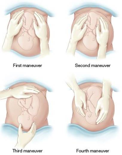
Leopold's Maneuver is a systematic abdominal palpation to estimate fetal size, locate fetal back and parts, and determine fetal position and presentation.
- Explain the procedure and its purpose.
- Position: dorsal recumbent with knees slightly flexed to relax the abdominal muscles.
- Draping
- Examining hands should be warm to avoid contractions of the abdominal muscles, which may impede palpation.
- Apply gentle, firm palpations with the palms of the hands.
The steps are divided into four:
- Palpate the fundus; check for breech or cephalic; usually breech— soft, globular, non-ballotable.
- Palpate the sides of the abdomen; check for a smooth, resistant back and irregular, small fetal parts of the fetus. The area of the fetal back is the best site for FHT auscultation.
- Palpate the area just above the symphysis pubis; check for cephalic or breech, usually cephalic; check the position and mobility of the head.
- Palpate the midline downward and just about two inches from the Pourpart’s ligaments; check for position and descent of the head (engagement), including degree of flexion (attitude).
Infobox
Leopold's Maneuver
Step Aliases
Step Name LM1 Fundal Grip LM2 Lateral Grip LM3 Pawlick’s Grip LM4 Pelvic Grip
Hygiene of Pregnancy
Nutrition
Always start with the dietary history when giving nutritional instructions to the mother. A Nutritional Profile should contain: pre-pregnant and current nutritional status, dietary habits (food type, quality, schedule, amount, culture), and the mother’s knowledge of nutritional needs.
Pica
Pica is a craving for the consumption of items not culturally defined as food. It is a common psychobehavioral disorder manifested during pregnancy, and can displace nutritious foods, interfere with nutrient absorption, and cause anemia.
- Common cravings include coffee grounds, clay, dirt, chalk, baby powder*, ice, charcoal, ash, eggshells, feces, hair, string, cloth, paint chips, laundry starch, paper, pet food, pebbles, and soap. (Geddes, 2023).
- Treatment is often not necessary; the disorder tends to resolve by itself. In cases of lingering pica, coping mechanisms or behavioral therapy sessions might be recommended.
Poor nutritional status may manifest with anemia, dull hair, dry/scaly skin, pale/dull mucus membranes/conjunctiva, and being under- or overweight. Particularly, the following the factors require special attention:
- Primigravidity
- Low pre-pregnant weight (<90 lbs.)
- Obesity
- Low socioeconomic status/economic deprivation
- Pre-pregnant debilitating conditions
- Vegetarianism, lacking essential protein and minerals and may need B12 supplementation.
- Successive short-interval pregnancies
- Education: nutritional teaching focusing on affordable but nutritious foods.
Calories
- Non-pregnant: 1,800 to 2,200 Kcal/day.
- Pregnant: +300 Kcal/day; 2,100 to 2,500 Kcal/day
- Avoid empty (non-nutritious) calories, such as calories gained from soft-drinks.
Essential Nutrients
- Protein: +30 g/day; 74 to 76 g/day. Milk, meat, fish, poultry, eggs.
- Carbohydrates: sufficient intake for energy needs (see calories), while avoiding empty calories.
- Fiber: fruits, vegetables; prevents constipation.
- Fats: energy-dense foods for absorption of fat-soluble vitamins (ADEK). Avoid too much fat to prevent vomiting and heartburn.
Minerals and Vitamins
- Iron: most important mineral; supplemented. It aids in the required increase in maternal RBC (~30%) and fetal liver storage in the third trimester (8th month).
- 18 mg/day (non-pregnant), and 30 to 60 mg/day (pregnant). Liver, read meats, green leafy vegetables, egg yolk, cereals, dried fruits, and nuts.
- Best absorbed in an acidic medium; take between meals and with Vitamin C-rich juice.
- Can contribute to constipation; adequate increase of fibers, fluid, and activity is also recommended.
- Darkened stool is normal.
- Calcium: needed for maternal calcium and phosphorus metabolism and fetal bone and skeletal growth (particularly in the 7th month).
- 1,200 mg/day, equivalent to 1 quart of milk a day (four glasses). Milk, milk products, and broccoli.
- Sodium: the most abundant cation in extracellular fluid. It effectively dictates fluid retention. Most foods contain sodium, and is not restricted unless seriously indicated. It is required for fetal tissue growth and development.
- Folic Acid/Folate/Vitamin B9: important for blood product production, metabolic demands in pregnancy, proper development of the fetus. Deficiency can result in fetal anomalies, neural defects, brain and spinal cord abnormalities, and bleeding complications.
- Neural Tube Defects (NTDs) are birth defects of the brain, spine or spinal cord that happen in fetuses within the first month of pregnancy, often even before awareness of the pregnancy. The two most common forms of NTDs are spina bifida and anencephaly.
- 0.8 mg/day (+100% requirement to prevent fetal malformations and neural tube defects). Liver, dark green leafy vegetables, avocados, papayas, and beans.
- More: legumes, asparagus, eggs, leafy greens, beets, citrus fruits, Brussels sprouts, broccoli, nuts and seeds, beef liver, wheat germ, papaya, bananas, avocado, and fortified grains. Fruits, green and leafy vegetables, nuts, and seeds, and fortified foods.
- Vitamins: water-soluble vitamins C and B and fat-soluble vitamins A, D, E, and K.
| Vitamin | Source |
|---|---|
| C | Citrus fruits and vegetables e.g. broccoli, bell peppers, and tomatoes |
| B Group | Legumes, beans, nutes, whole grain, oatmeal, pork, beef, fish, liver, organ meats, eggs, and green leafy vegetables |
| A | Milk and dairy products, dark green and dark yellow fruits and vegetables, eggs, and liver |
| D | Milk and foods fortified with vitamin D, egg yolk, fish |
| E | Nuts, seeds, wheat germ, whole grain products, green leafy vegetables, vegetable oils |
| K | Meats, liver, cheese, tomatoes, peaks, egg yolk |
| Food | Number of Servings |
|---|---|
| Milk and milk products | 1 quart (4 glasses) a day |
| Meat and meat products | 3 to 4 servings |
| Cereals/grain products | 4 to 5 servings |
| Fruit/fruit juices | 3 to 4 servings with at least one serving of vitamin C-rich fruit/juice |
| Vegetables/vegetable juices | 3 to 4 servings with at least one serving of dark green or yellow vegetable |
| Fluids | 4 to 6 glasses of water plus other fluids to equal 8 to 10 cups/day |
| The WHO also suggests two priority antenatal nutrition recommendations: Multiple Micronutrient Supplements (MMS, often contains 30 mg; 60 mg if anaemia is a severe public health problem) and Vitamin D supplements during pregnancy. |
Bath and Clothing
A daily bath can be taken if desired, but soap should not be used for cleaning the nipples; they produce a drying effect. The nipples should be dried with a towel to increase their toughness/integrity.
- Tub baths should only be done with care and anti-slip precautions (nonskid rubber mat). The risk of falls is great with the loss of balance in pregnancy.
- Douching, especially for leukorrhea associated with elevated estrogen levels, is not necessary. A daily bath should suffice.
Clothing should be comfortable and loose; not constricting especially around the breasts, abdomen, or legs. Round garters should not be used.
- Flat-heeled shoes help maintain balance and comfort.
- Supportive hip-high panty hose is used to manage varicosities.
- Supportive, cotton-lined brassieres may be used.
- Maternity girdle can be used as necessary.
Sleep and Rest
- Assess activities to identify the need for rest and sleep.
- The average number of hours of sleep is 8. 1 to 2 hours of afternoon nap may be required. In the second half of pregnancy onwards, sleeping supine should be advised against.
- Plan rest times during the day.
- At work, get to stand and walk for a few minutes at least once every 2 hours (if the task requires prolonged sitting, there should be time to walk about and sit in intervals)
Traveling
- Long distance travelling should provide stopovers so pregnant women can get out of the car and walk. Seatbelts are needed.
- Air travel before the 36th week of pregnancy is considered safe, but with prior checking with the health care provider. Some flights require a medical certificate indicating fitness to travel by air.
- The best time for travel is the second trimester, when the woman feels most comfortable, with the least risk for abortion and premature labor.
- Aisle seating in various forms of transportation is ideal.
- Journeys when close to term is discouraged.
Exercises
- Generally, low-impact and moderate intensity exercises are done. These commonly include walking progressing to jogging, stationary cycling, routine swimming, and yoga.
- Cleansing breathing: deep, relaxed breathing (i.e., a sigh). Can be practiced in pregnancy and used in labor to signal the beginning of uterine contractions.
- Pelvic Rock: highly important for comfort, this exercise increases the flexibility of the lower back, strengthens the abdominal muscles, shifts the center of gravity back to the uterine spine, and relieves backache and improves posture and appearance in late pregnancy.
- Squatting or Tailor-sitting: strengthens the perineal muscles, making pelvic joints more pliable.
- Abdominal Breathing: primarily utilizing the diaphragm to breath rather than the chest muscles, helpful during the first half of labor. With total relaxation, it can carry women through most of the first stage.
- Kegel: improves the tone of pubococcygeal, perineal, vaginal, and pelvic flor muscles needed for pregnancy, labor, and delivery, in uterine prolapse, cystocele, and rectocele; this can be done hourly.
- Panting: best for the crowning period, leaving the work to be accomplished by the uterus. Only by panting can the mother avoid pushing in the transition phase of labor; pushing should be in the second stage of labor except during crowning.
Remember to avoid dehydration, fatigue, contact sports and , high-impact and high-intensity or excessive/strenuous exercises. Maternal exercises do not need to be limited as long as usual and customary and does not cause fatigue. Exercises reduce the rates of emergent cesarean section rate and length of hospitalization. Exercises should be stopped if danger signs are present: dizziness, headache, overheating, fast and unsteady heart rate, difficulty breathing, and nausea; seek emergency management if vaginal bleeding, uterine contractions, abdominal or pelvic pain, or chest pain occurs.
Marital Relations and Coitus
The gravid woman loses interest in sexual intercourse during the first trimester due to fatigue, nausea, or adaptation to pregnancy; and the third trimester due to discomforts of pregnancy. The second trimester may feature regained interest in sex as it is the most comfortable period of pregnancy.
Generally, no restricts are placed except in the presence of premature rupture of membranes (PROM), premature labor, history of abortion and bleeding, deep engagement of the head in late pregnancy, and an incompetent cervix. In healthy pregnant women, sexual intercourse does not usually cause harm. Avoid fatigue, exercise moderation. It is suggested to take on side-lying and woman-on-top positions.
Employment
Work may continue, provided it does not pose hazards to the health of the mother and fetus. Safety and rest are the two most important considerations in deciding whether or not the pregnant woman should stop working.
- The mother should be able to walk every few hours to improve blood circulation, regardless of sitting or standing during work.
- Adequate rest periods should be provided.
- Previous conditions, which are likely to be repetitive (e.g., SGA, premature labor, abortions) should minimize physical work.
- Maternity leave: R.A. 7322 amended by R.A. 11210, the Expanded Maternity Leave Act.
| Republic Act 7322 | Republic Act 11210 |
|---|---|
| 60 days of maternal leave for NSD and 78 days for CS 15 days of paternity leave | 105 days paid leave between prenatal and postnatal leave, at least 60 days of which should be for the postnatal period. + 15 days for parents qualifying as a solo parent under R.A. 8972 + Optional additional 30 days of unpaid leave. |
| For the first four pregnancies only | No limit on applicable pregnancies regardless of mode of delivery |
| In case of an emergency termination or miscarriage, 60 days of unpaid leave is provided. |
Oral Hygiene
The teeth and gums should be regularly examined as part of the prenatal general physical examination. Dental caries require prompt management in pregnancy, but major dental surgeries should be postponed for the postpartal period.
- Due to estrogen’s effect on vascularity, the gums of pregnant women are painful and swollen. Instruct them on the use of a soft-bristled toothbrush and gentle brushing.
- The concept that dental caries are aggravated by pregnancy is not supported by literature; there is no tooth loss secondary to pregnancy.
SAD Habits
- Smoking: should absolutely avoided, whether cigarettes or electronic cigarettes. This also applies to secondhand smoke.
- Smoking is a risk factor for SGA, prematurity, infant mortality, spontaneous abortion, placenta previa, abruption placenta, and premature rupture of membranes. It also causes learning and attention problems in children.
- This is caused by the vasodilator effect of nicotine on the body, reducing placental perfusion. Carbon monoxide binds to hemoglobin, inactivating maternal and fetal hemoglobin. Smokers also have decreased plasma volume and reduced appetite.
Caffeine
Caffeine also produces vasoconstriction, and is worse when combined with alcohol. The WHO recommends a caffeine limitation of 300 mg per day, nor more than 2 to 3 servings.
- Alcohol: alcohol ingestion is likely to cause fetal abnormalities. It is the leading known teratogen in the west. There is no safe amount of alcohol in pregnancy.
- Alcohol also produces pronounced learning and attention problems, more consistently than smoking does.
- Fetal alcohol syndrome (FAS) occurs at a rate of 10% of births with heavy use of alcohol (2 or more drinks per day). FAS is characterized by: retardation (cognitive, motor, attention, learning), microcephaly, seizure disorders, craniofacial defects (“FAS facies”; flat midface, wide nasal bridge, thin upper lip), cardiovascular defects, limb defects, and impaired fine and gross motor function.
- Drugs should only be taken if prescribed by their physician. Risks should never outweigh the benefits of taking drugs. Illicit drugs can result in the most adverse fetal malformations in the first trimester, as (1) the placental barrier is not yet fully developed until the 10th to 12th week of gestation, and (2) rapid organogenesis occurs within the first two months of pregnancy.
- “Hard drugs” may cause growth retardation and drug withdrawal, which are associated with increased neonatal mortality. The most common harmful effect of heroin on neonates is withdrawal or neonatal abstinence syndrome, giving rise to a group of signs that include sneezing, irritability, vomiting and diarrhea, and seizures.
- The use of illegal drugs also come with the risk of acquiring HIV and other STDs because women may trade sex for drugs, or provide sexual favors for the money to acquire drugs.
- Herbal drugs are not always safe. As a general rule, they must be approved and supervised by the health care provider.