Oxygenation is the distribution of oxygen to the body’s cells via internal and external respiration.
Heart
A cone-shaped, hollow, muscular organ location in the mediastinum between the lungs. It pumps ~5L/min (Cardiac Output) or ~60mL/beat (Stroke Volume).
- It is covered by a thin covering called the pericardium, made up of the parietal pericardium, the visceral pericardium, and the pericardial cavity/space that sits in between them, containing pericardial fluid normally around ~20mL.
- There are three layers of cardiac muscle tissue: the epicardium, myocardium, and endocardium (from outermost to innermost).
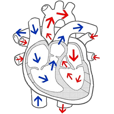
Chambers and Valves of the Heart
- Right Atrium: entry point of unoxygenated blood into the heart from the SVC, IVC, and Coronary Sinuses.
- Exit Point: tricuspid valve (when closing, it produces S1)
- Right Ventricle: exits into pulmonary circulation.
- Exit Point: pulmonary semilunar valve (when closing, produces S2/P2)
- Left Atrium: entry point of oxygenated blood into the heart from the four pulmonary veins.
- Exit Point: mitral/bicuspid valve
- Left Ventricle: exits into systemic circulation.
- Exit Point: aortic semilunar valve (when closing, produces S2/A2)
Coronary Artery Disease
The coronary arteries are the main oxygen supply of the heart. When they are blocked by plaque formed from deposition and damage caused by fat/lipids, atherosclerosis occurs, which leads to angina pectoris, where the oxygen supply and oxygen demand of the heart is mismatched. This leads to necrosis, which leads to myocardial infarction.
- Left Coronary Arteries
- Left Anterior Descending Artery (LADA): supplies the LV and Ventricular Septum, Chordae Tendinea, Papillary Muscle, and to a lesser extent, the RV.
- Circumflex Coronary Artery: supplies the LA, Lateral and Posterior Surfaces of the LV, Some of the Ventricular Septum, and the SA/AV Nodes.
- Right Coronary Artery: supplies the RA, RV, and the lower portion of the LV.
Electrophysiologic Properties of the Heart
- Automaticity: spontaneous and automatous repetitive impulse.
- Excitability: depolarization, the response to a stimulus.
- Conductivity: transmission of impulses across the heart.
- Contractility: physical contractile ability of the heart.
- Refractoriness: inability to respond until repolarization.
- Elasticity/Distensibility: ability of the heart to compensate increased pressure and volume.
- Rhythmicity: standard and continuous repolarization and depolarization of the heart.
Conduction System of the Heart
- Sinoatrial Node: the pacemaker of the heart (because it’s the fastest), which sends impulses at around 60 to 100 BPM, the normal adult heart rate.
- Sends impulses to the two atria (via the internodal tracts) initiating their contractions (P Wave).
- The impulse also reaches the AV Node, where the impulse is delayed to wait for the atria to finish contracting.
- Atrioventricular Node: secondary pacemaker if the SA Node fails, sending impulses at around 40 to 60 BPM.
- Bundle of His
- Right and Left Bundle Branches
- Purkinje Fibers: ends in ventricular depolarization (QRS Complex)
NOTE
Disturbances in the conduction system of the heart is called “Heart Block”.
Mechanical Properties of the Heart
- Cardiac Output: the product of an individual’s heart rate and stroke volume ().
- Increased by stimulation of the autonomous nervous system and increased release of endogenous catecholamines (epinephrine, norepinephrine, dopamine).
- Decreased by the parasympathetic nervous system (vagus nerve), Beta-Adrenergic Blockers (-olols), Calcium Channel Blockers (-dipines), Digitalis (Digoxin).
- Stroke Volume: amount of blood pumped in one beat of the heart.
- Preload: the amount of blood distending the ventricles just before contraction.
- Afterload: the resistance the ventricles must overcome to eject blood.
- Contractility
Vascular System
Hmm 🤔